Urogenital Chlamydia is an infection that is transmitted in most cases sexually, the causative agent of which is Chlamydia trachomatis - a gram-negative intracellular bacterium belonging to the order Chlamydiales, family Chlamydiaceae, genus Chlamydia.
Serotypes C. trachomatis A, B, Ba, C, are the causative agents of trachoma; D-K - urogenital Chlamydia; L1, L2, L3 - lymphogranuloma venereum.
In the human body, chlamydiae are in two forms, which differ in structural and biological properties:
- highly infectious - (spore-like, extracellular, which does not multiply and is not sensitive to antibiotics) form - is an elementary body (ET)
- vegetative -- (which multiplies, intracellular, sensitive to antibiotics) form - is the reticular body (RT).
At the initial stage after infection, ET is fixed on the host cell plasmalemma, followed by the penetration of Chlamydia into the cell by endocytosis. During the first 7-10 hours, the intussusception of the plasmalemma site with adsorbed ET into the cytoplasm occurs with the formation of a phagocytic vacuole, where further within 6-8 hours ET turns into a vegetative form - RT, capable of growth and further reproduction.
Reproduction of Chlamydia leads to the formation of special inclusions (Gelberstedter's bodies - Provachek). In the first 18-24 hours of development, they in the amount of 100 to 500 chlamydiae are localized in the cytoplasmic vesicle, which consists of the cell membrane. Later, within 36-42 hours, the process of maturation of the RT occurs through the transitional (intermediate bodies) (PT) and the development of ET of the next generation. The full cycle of reproduction of Chlamydia lasts 48-72 hours and ends with the death of the cell affected by Chlamydia; however, under adverse metabolic conditions for the vital activity of Chlamydia, this process can take time. Reproduction of Chlamydia in epithelial cells leads to disruption of the integrity of the epithelial layer, its desquamation, as well as lymphoid tissue infiltration.
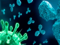
In addition, chlamydiae have the ability to suppress the fusion mechanism of phagosomes with lysosomes, followed by inhibition of the growth of Chlamydia in monocytes and macrophages in an intermediate state at the stage between ET and RT and the formation of so-called "aberrant" RTs, which produce a minimal amount of structural antigens, but strongly express chlamydial heat shock protein - HSP-60, which has a high similarity with the same protein of the host cell membrane, as a result of its immune and phagocytic systems are unable to recognize this antigen as foreign, and therefore antibodies to it can also cause autoimmune tissue damage, through increased synthesis of autoantibodies, including antisperm, leading to immunological infertility.
The resistance of non-developing RT to antichlamydial antibiotics is associated with the cessation of their metabolic processes, as well as with the causative agent's experience of the period of drug therapy when they are in special membrane-protected zones of the epithelium, Trichomonas, as well as in neutrophilic leukocytes, macrophages, lymphocytes, in endothelial capillaries, lymphatic extracellular phagosomes. Thus, persistent chlamydial infection is an autoimmune process that contributes to the maintenance of chronic inflammatory processes and does not require the presence of a microbial agent in its further course.
Chlamydia affects the cylindrical epithelium of the urethra, cervical canal, conjunctiva of the eyes and rectum, and occasionally the oropharynx. In adult women, Chlamydia is not able to vegetate in the unchanged stratified squamous epithelium of the vagina due to the high sensitivity to the acidic environment of its contents. Therefore, the primary lesion is usually the mucous membrane of the cervix of the cervical canal. In childhood and adolescence, chlamydia, on the contrary, can cause damage to the mucous membranes of the vulva and vagina. This is facilitated by the anatomical and physiological features of the reproductive system of girls (incomplete physiological defence mechanisms, a small number of layers of superficial epithelium, alkaline PH of vaginal discharge).
Chlamydia is also sometimes absorbed by peripheral monocytes and spreads throughout the body, settling in various tissues, turning into tissue macrophages, which can remain viable for several months while being an antigenic stimulant, promoting the formation of fibrous granulomas in healthy tissue. Infection of adults with Chlamydia occurs sexually through contact with patients with Chlamydia, children - by the perinatal route (antenatal - through the placenta, intrapartum - during childbirth) and through sexual contact; less often, from sick mothers to children, more often women, even less often young girls can become infected if the rules of personal hygiene and child care are violated (contact - household way). The risk of infection in women increases when using oral contraceptives due to the loss of resistance of epithelial cells to this infection due to changes in hormonal levels, as well as during abortions and other surgical interventions on the organs of the genitourinary system. With HIV infection, people are more prone to contracting Chlamydia, and urogenital Chlamydia, in turn, increases susceptibility to HIV-1 infection, and HIV strains isolated from such patients are more virulent.
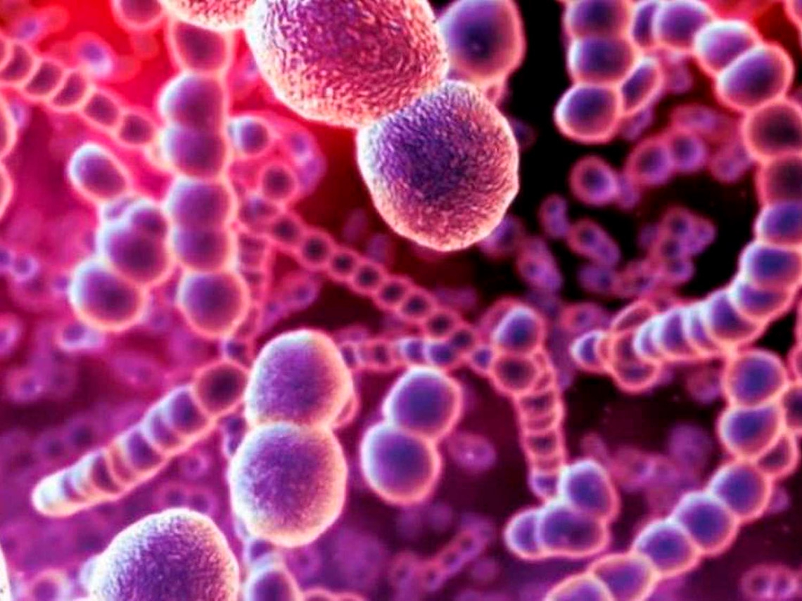
Urogenital chlamydia is a very common STI. The WHO estimates that 131 million people are infected with C. trachomatis every year, with the highest incidence in those under the age of 30. Chlamydia is currently ranked 2nd among STIs after trichomoniasis, ahead of gonorrhoea and syphilis. Chlamydia is very often associated with other STIs and opportunistic pathogens. Mixed infections, in turn, increase the pathogenicity of each microorganism included in the association, affect the incubation period and the clinical course of the inflammatory process (the most striking clinic is characteristic of the association with trichomoniasis and gonorrhoea), complicate their diagnosis and especially treatment, contribute to their chronicity and recurrence ...
Chlamydia as a mono-infection is extremely rare, only in 2%. In women with inflammatory diseases of the genitourinary organs, the association of Chlamydia and gonorrhoea reaches 30%, associations with Trichomonas, a conditionally pathogenic flora, are also very frequent(ureamicoplasma, candida, Gardnerella, viral infections (HSV, HPV), Escherichia coli, staphylococci and enterococci). Mixed infection is also characterized by multifocal lesions (both urogenital and extragenital), difficulties with both accurate diagnosis and subsequent treatment.
Classification of diseases caused by the presence of C. trachomatis
- Chlamydia of the lower genitourinary system: urethritis, cervicitis, cystitis, vulvovaginitis.
- Chlamydia of the pelvic organs and other organs of the genitourinary system: paraurethritis, epididymitis, orchitis, prostatitis accompanying urethritis (in men); vestibulitis, salpingo-oophoritis, endometritis, pelvic peritonitis (in women). a) Chlamydia of the genitourinary system, not exactly localized.
- Chlamydia of the anorectal region.
- Chlamydial pharyngitis, tubo-otitis, otitis media.
- Chlamydia of extragenital localization (arthritis, pneumonia, perihepatitis).
- Chlamydial conjunctivitis.
Chlamydia of the lower genitourinary system
in women, it manifests itself in the form:
- swelling and redness of the mucous membrane of the external opening of the urethra
- infiltration of the walls of the urethra
- Abundant discharge of mucus and pus from the urethra
- swelling and redness of the mucous membrane of the cervix
- discharge of mucus and pus from the cervical canal
- erosion and dysplasia of the mucous membrane of the cervix (in association with other STIs).
in men:
- swelling and redness of the mucous membrane of the external opening of the urethra
- infiltration of the walls of the urethra
- insignificant discharge of mucus and pus from the urethra.
Chlamydial pharyngitis:
- In women and men, in most cases, there is a clinically asymptomatic course of the disease. In the presence of clinical manifestations, subjective symptoms may disturb: dryness in the oropharynx; pain that worsens when swallowing; sometimes combined with tubo-otitis and otitis media
- on the examination - swelling and redness of the mucous membrane of the oropharynx and tonsils
Chlamydial conjunctivitis - clinically occurs in the form of simple or follicular conjunctivitis in acute, subacute or chronic forms with complaints of:
- dryness, photophobia, redness and swelling of the conjunctiva of the affected eye
- slight soreness of the affected eye
- slight discharge in the form of mucus and pus in the corners of the affected eye.
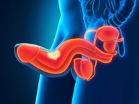
Chlamydia of the pelvic organs and other organs of the genitourinary system
in women:
more than 70% of women are characterized by an asymptomatic course of the disease. In the presence of clinical manifestations, subjective symptoms disturb:
- vestibulitis (slight or moderate secretion of mucus and pus from the genital tract, redness of the external openings of the ducts of the vestibular glands, their soreness and swelling)
- salpingo-oophoritis (for the acute course of the inflammatory process - pain in the lower abdomen of a cramping character, discharge of mucus and pus from the genital tract and cervical canal is characteristic; for the chronic course of the disease - clinical manifestations are less pronounced, characterized by a violation of the menstrual cycle; in acute course - increased , painful on palpation of the fallopian tubes and ovaries, shortening of the vaginal vaults; in the chronic course of the disease - slight pain, compaction of the fallopian tubes, their limited mobility on palpation)
- endometritis - pain in the lower abdomen, usually of a pulling nature (for the acute course of the disease, a painful, enlarged uterus of soft consistency, mucus and pus discharge from the cervical canal is characteristic; for the chronic course of the disease, subjective manifestations are less pronounced, post- and intermenstrual minor spotting; on palpation - a dense consistency and limited mobility of the uterus)
- pelvioperitonitis (characteristic appearance - general serious condition, hectic body temperature, hypotension, nausea, vomiting, oliguria, sharp abdominal pain on superficial palpation, in the lower sections, the tension of the muscles of the abdominal wall and a positive symptom of irritation of the peritoneum, impaired defecation are determined).
in men:
- epididymis-orchitis (typical: discharge of mucus and pus from the urethra; palpation determines an enlarged, dense and painful testicle and its epididymis; soreness in the epididymis and groin, often one-sided; pain in the perineum radiating to the perineum, to the lower part abdomen, in the scrotum, on the spermatic cord, inguinal canal, lumbar region, sacrum; there are hyperemia and swelling of the scrotum in the affected area)
- prostatitis accompanying chronic urethritis - pain in the perineum and in the lower abdomen radiating to the rectum, dysuria (on palpation, a painful, compacted, sometimes enlarged, and edematous prostate gland is determined).
In men and women, Chlamydia of the paraurethral glands is possible, with manifestations in the form of itching, burning, pain during urination (dysuria); with a characteristic discharge of mucus and pus from the urethra; the presence of dense painful formations, the size of millet grain, in the area of the excretory ducts of the paraurethral glands; pain in the area of the external opening of the urethra; pain during intercourse (dyspareunia).
Clinical symptoms of childhood chlamydia are similar to clinical symptoms in adults, with more pronounced and vivid clinical symptoms of lesions of the mucous membranes of the vulva and vagina in girls.
Chlamydial STIs, non-sexual localization
Reactive chlamydial arthritis (disease, Reiter's syndrome) is characterized by a course in the form of urethrooculosinovial syndrome, which classically manifests itself as a triad (complete or incomplete): urethritis, conjunctivitis, arthritis. Reiter's syndrome can also be accompanied by lesions of the skin and mucous membranes (keratoderma, circinar or xerotic balanoposthitis, ulceration of the oral mucosa), as well as symptoms of damage to internal organs(cardiovascular, nervous system and kidney). In 80-95% of cases, Reiter's disease is associated with the HLA-B-27 antigen. The presence of a common antigenic determinant in the causative agents of Reiter's disease and HLA antigens leads to insufficient elimination of microorganisms, the emergence of latent infection and the formation of an autoimmune process due to the cross-reaction of HLA-B-27 with hsp60 Chlamydia.
With reactive arthritis, the following joints are affected: knee, ankle, metatarsophalangeal, toes, hip, shoulder, elbow, and others. The disease often occurs in the form of polyarthritis (on average, 5-6 joints are affected). There are two stages of Reiter's disease: infectious-toxic(with a therapeutic effect from anti-Chlamydia antibiotics) and autoimmune (when the elimination of the infectious factor does not significantly affect the course of the disease, in which persistent or recurrent foci of immune inflammation in the joints or other organs are formed).
The average duration of the first stage of the disease is 4-6 months. For reactive arthritis, a wave-like course is characteristic: in 50% of cases, relapses of the disease are observed at various intervals. In 20% of patients, various muscle atrophies and enthesopathy are revealed, with the most frequent lesions of the Achilles tendon and plantar fascia, which causes gait disorders.
With disseminated Chlamydia in men and women, pneumonia, perihepatitis, peritonitis can develop.
Chlamydia of the anorectal region
In men and women, there is usually a clinically asymptomatic course of the disease. In the presence of clinical manifestations, the following clinical symptoms are characteristic: with local lesions of the rectum: itching, burning in the anorectal region, slight discharge from the rectum of a yellowish-pink colour; when the process is localized above the anus: painful tenesmus, soreness during bowel movements, mucopurulent discharge from the rectum, often mixed with blood.
Laboratory diagnostics:
The diagnosis of Chlamydia is established based on the results of laboratory studies using molecular biological methods aimed at detecting specific fragments of DNA and/or RNA of C. trachomatis (such as PCR, real-time PCR or NASBA), depending on the localization of the infectious process in the body, as well as data sexual history: smears from the mucous membrane of the oropharynx, discharge from the mucous membrane of the rectum, mucous membranes of female genital organs, from the urethra, semen, prostate secretions, conjunctiva, examination of the first portion of urine for C. trachomatis.
Examination for Chlamydia is mandatory if other STIs are detected (especially after-treatment of the often accompanying trichomoniasis and gonorrhoea) and opportunistic flora (gardnerellosis, mycoureaplasmosis), viral (HSV, HPV), candidiasis, staphylococci and enterococci, Escherichia coli or dysplasia, the presence of erosion cervix, inflammatory types of urogenital smears, chronic prostatitis, infertility (both female and male or combined), not carrying a pregnancy up to 12 weeks (frozen or undeveloped pregnancy), a history of ectopic pregnancy, after unprotected sexual intercourse.
The sensitivity of molecular biological methods is estimated at 98-100%, specificity - 100%. It is strictly necessary to comply with the conditions for transporting clinical material to the laboratory.
False-positive PCR results are the result of so-called cross-contamination with amplification products. False-negative results occur due to the presence of substances in the clinical material that inhibits PCR.
More rarely, methods such as are used to diagnose Chlamydia:
- cultural - but due to laboriousness and duration, problems with reproducibility, the need for expensive equipment, media, instruments, dishes, qualified personnel - is rarely used in routine clinical practice, but only in scientific research
- cytological - but due to low sensitivity, laboriousness and high subjectivity, this method is practically not used
- enzyme-linked immunosorbent assay (ELISA) - determination of immunoglobulins of classes: M, G, A.
- immunohistological method of luminescent antibodies: direct (PIF), an indirect immunofluorescence method (NPIF) - screening methods. False-negative results may be related to a small number or absence of epithelial cells in the smear, and false-positive results - as a result of nonspecific binding of antibodies to uninfected material or other Chlamydia species, as well as insufficient purification of monoclonal antibodies - nonspecific fluorescence.
- immunochromatographic and enzyme-specific - indirect (screening) tests for laboratory diagnosis of urogenital Chlamydia.
Healing control:
- sampling of material for control studies should be carried out no earlier than one month (for all PCR modifications) and 14 days (for NASBA) after the end of the course of antibiotic therapy with drugs active against C. trachomatis. The sensitivity of the study can be influenced by various inhibiting factors, as a result of which strict requirements are imposed on the organization and operation of the laboratory in order to exclude contamination of clinical material and to minimize both false-positive and false-negative results.
The use of laboratory methods other than molecular biological, such as the method of direct immunofluorescence (DIF), enzyme-linked immunosorbent assay (ELISA) for the detection of antibodies to C. trachomatis, microscopic and morphological methods for the diagnosis of Chlamydia - not currently recommended due to their lower sensitivity and specificity than PCR with more false-positive and false-negative results. The use of biological, chemical and alimentary provocations in order to increase the efficiency of diagnosis and in the treatment of chlamydial infection is not recommended.
To exclude inflammatory diseases of the pelvic organs and the complicated course of Chlamydia, it is recommended to conduct ultrasound examination of the pelvic organs (both transabdominal and transvaginal and transrectal), and, if necessary, CT or MRI with contrast enhancement).
Consultation of related specialist doctors is recommended, such as a urologist, gynaecologist, ophthalmologist, otorhinolaryngologist, proctologist, rheumatologist, in children - neonatologist, paediatrician - in order to clarify the scope and nature of the additional examination.
Conservative treatment
Recommended for the treatment of Chlamydia of the lower genitourinary system, anorectal region, chlamydial pharyngitis, chlamydial conjunctivitis, oral administration of three groups of antibiotics:
- Macrolides: azithromycin, josamycin, clarithromycin.
- Fluoroquinolones: ofloxacin, levofloxacin
- doxycycline
The duration of the course of treatment is strictly individual and depends on the degree of clinical manifestations of the inflammatory processes of the genitourinary organs, the results of laboratory and instrumental studies, the presence of often concomitant other STIs. Depending on the above factors, the duration of therapy can vary from 10-14 to 21 days, in the presence of Reiter's disease from 4 to 7 weeks. It is important to note that the use of insufficient doses of drugs to cure can induce the formation of persistent forms and L-forms of the pathogen of Chlamydia.
Treatment of pregnant women with Chlamydia is carried out at any stage of pregnancy with antibacterial drugs, taking into account their effect on the fetus with the participation of obstetricians - gynaecologists. Treatment of Chlamydia in children weighing more than 45 kg is carried out in accordance with the prescription regimens for adults, taking into account possible contraindications.
For the treatment of children weighing up to 45 kg, it is recommended to prescribe azithromycin per os, based on a dose of 10 mg per kg of body weight for at least 7-10 days.
Prevention:
- exclusion of casual sex;
- use of barrier contraception, thorough toilet of the external genitalia with the use of antiseptics;
- examination and treatment of sexual partners;
- screening for other STIs;
- in case of an unknown source of infection, repeated testing for syphilis is recommended after three months, for HIV, hepatitis B and C - after 3-6-9 months.
In the absence of the effect of treatment, it is recommended to exclude both reinfection and prescription of drugs of a different pharmacological group and treatment, taking into account the often concomitant other