Determination of the echostructure of the prostate by the method of bridles through the anterior abdominal wall, as a rule, is impossible due to the significant distance of this organ from the scanning surface of the sensor, shielding part of the organ by the pelvic bone structures. Minor changes in the structure of the organ, characteristic of the early stages of cancer, are not accompanied by changes in the shape, the symmetry of the organ, violation of the integrity of the structure of the capsule, are not registered because they are outside the resolution of the method. Localized cancer is usually not detected by a transabdominal examination. Removal of local spread in the transabdominal examination is possible only with the spread of tumors in the vascular organs, which gives preconditions in the process of abuse. In most cases, even in large glands with pronounced asymmetry, echostructure disorders, with hypoechoic combined structures, differentiated BPH and cancer, not detected as possible. In TRUS, the surface of the sensor is removed from the object of study only by a thin wall of the rectum, Denoneville aponeurosis, a thin layer of fat in 2-6 mm.
High-frequency (5-16 MHz) sensors with high-resolution properties are used. Besides, the advantages of the method include the lack of need to fill the bladder, which is sometimes impossible in patients with urinary incontinence or irritable symptoms. Although it should be noted that even a small filling of the bladder can help in TRUS to detect even slight germination of prostate cancer in the bladder or primary bladder cancer. The preclinical stage of prostate cancer diagnostic is possible during TRUS.
The minimum tumor size detected in TRUS is 5 mm, which allows detecting tumors of small volume, located in the depth of the organ and inaccessible during a rectal examination. However, a large number of tumors are isoechoic to prostate tissue, so TRUS is normal gray-scale mode has a low sensitivity in the range of 57-70% in the detection of prostate cancer in asymptomatic men. It is known that prostate cancer in 70-80% of cases is the result of the degeneration of cells in the peripheral zone; the primary tumor node is usually located at a distance of 3-4 mm from the capsule of the prostate. Less than 5% of prostate cancers originate from the central zone. Most cancers identified in this area spread here from the peripheral area, and in 25% of cases in the area of "anatomical weakness" - in the course of the ejaculatory ducts is the invasion of the seminal vesicles. Other areas of "anatomical weakness" are the base and apex of the gland, which do not have a capsule. Transitional areas make up only 5% of the total glandular mass of the prostate. However, in 10-20% of cases, prostate cancer is detected in them. Since the transition zones are the site of almost exclusively benign hyperplasia. The incidence of cancer in TRUS in the transition areas is small and is only 19%. It follows that only 1 in 6 suspected foci of cancer in the transition zone will be a cancerous node, the rest - areas of microglandular hyperplasia, glandular atrophy, or a focus of inflammation in exacerbation of prostatitis. Histological diversity of tissues and tumors of the prostate leads to problems in the interpretation of the image obtained by TRUS. Even with normal glandular cancer from the peripheral zone, different echogenicity of tumors is observed depending on histological differentiation, age, and tumor volume. In 60% of cases, the tumor nodes look hypoechoic, in 39% of cases, the tumors are isoechoic to the sensory normal tissues, and, consequently, are not visualized. Hyperechogenic cancer was detected in 1% of cases. The hypoechogenic structure was provided by low-differentiated tumors. Highly differentiated tumors are usually isoechoic. In addition to the pathomorphology of the tumor, the age of the tumor is also important in creating a certain echostructure. Thus, the primary cancer node, most often found in the form of a hypoechoic rounded zone with a fuzzy contour in the peripheral zone, changes its echostructure and shape over time during its growth. This is due to both the age of the tumor and its ability to infiltrate glandular tissue (eg, hyperplasia of central glandular tissue). The growth of the primary node can occur outside, with subcapsular spread, with deformation of the contour and without it, as well as inside the gland with deformation of the surgical capsule, with the invasion of the central parts of the gland. The primary cancer node can be avascular, hypervascular, moderately vascular. With the growth of the node changes its echogenicity, structure, vascularization. The most important thing that should stand out, and what you need to pay attention to - is a violation of the normal ultrasound and vascular architecture of the gland. For example deformation of the surgical capsule with the spread of the tumor inside, to the central departments, the appearance of signs of the pathological vascular network, deformation of the rectal or posterolateral, lateral contour of the gland, rarely - the anterior contour; lack of differentiation of the peripheral zone and internal parts of the gland with the involvement of transitional zones; violation of vascular architecture in the form of abnormal location and course of blood vessels (appearance of vascular elements where they are not normally visualized), violation of blood flow symmetry, uneven vascular thickness and their pathological curvature, vascular diversity, vascular trifurcations. Over time and as the tumor ages, small foci of necrosis are formed, followed by fibrosis and calcification. At the same time, the primary hypoechoic structure of a tumor is lost, the tumor becomes inhomogeneous and can be undiagnosed, especially against the background of available nodes of an adenoma or against the background of the changes characteristic of chronic prostatitis. Massive accumulation of calcifications and areas of fibrosis should be treated with great caution, as massive calcification can mask the tumor process. Since 85% of prostate cancer is multifocal, often a biopsy from suspicious areas of the prostate can be negative, and a biopsy from a tissue that looks healthy - positive. At a biopsy of hypoechoic zones, detected in TRUS, 2 times more likely to be cancer, compared with pieces of tissue from isoechoic areas, so from 25 to 50% of cancers will be missed, if only it is carried out from hypoechoic areas.
Because of this, the search for methods to increase the informativeness of TRUS continues. The greatest hopes are placed in the following areas:
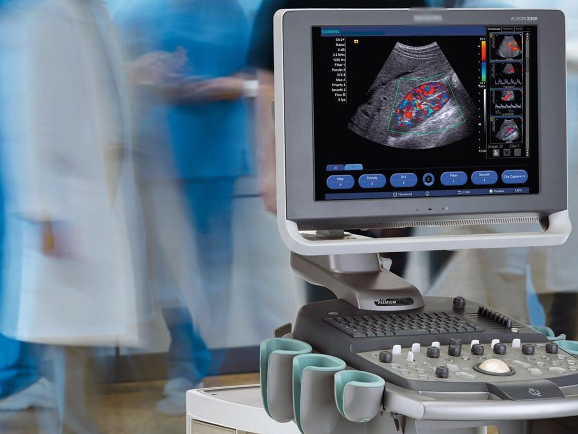
- Ultrasound Doppler in color-coded modes (changes in vascular architecture in the tumor is accompanied by the asymmetry of the vascular pattern in the lobes, the emergence of new tortuous vessels of different calibers, going in unusual, non-radial directions, the appearance of vascular trifurcations (vascular anastomoses), penetrating the capsule, and those leading to the tumor of the vessels. Thus, Doppler supplements the grayscale TRUS. However, the homogeneity of changes in the macrostructure of the gland in other diseases (infiltration, fibrosis, nodules, calcifications, cysts), as well as frequent combinations of diseases - cancer on the background of BPH, cancer on the background of chronic prostatitis, BPH and chronic prostatitis complicate diagnosis.
- Echocontrast studies - the use of liquids, which include air microbubbles, gas-containing microspheres, suspensions, which include gas microbubbles (echo contrast drugs) (EKD). EKD can be used in 2 basic modes: in the Doppler mode and the mode of the grayscale harmonic image. When exposed to the smallest vessels of the tumor with the energy of Doppler radiation, microbubbles burst and generate a powerful Doppler signal. In this case, microvessels that are not available for visualization may become visible. Harmonious images allow the use of contrast agents in the grayscale low-energy mode. The use of IVF helps to significantly increase the sensitivity of ultrasound in the detection of cancer. The main advantage of using IVF is the ability to diagnose the most aggressive, fast-growing forms of cancer.
- Ultrasound elastography. A new method for assessing the elasticity of the fabric (elasticity, compressibility, stiffness). Elasticity can change under the influence of various pathological processes, inflammation, tumor, fibrosis, atrophy. Elastography is performed in real-time with grayscale or color-coding. In color mechanical elastography, less elastic fabrics are mapped in blue, fabrics of medium elasticity are mapped in green, and elastic fabrics are mapped in red. Tumor tissue is usually more stiff or rigid and glows blue or cyan. The disadvantage of this method is the difficulty of uniform compression by the sensor, even taking into account the compression scales. This is especially true for the use of a rectal transducer, due to its design features when the vector of mechanical pressure exerted by the operator must correspond to the scanning angle. The technique is operator-dependent, with great difficulty in reproducing the results due to the lack of standardized assessment of the degree of compression, as well as standardized elasticity.
- The best qualitative information about the structure of the gland, close to the anatomical, is given by high-field MRI of 1.5 and 3 T with bolus contrast enhancement, which gives the best image. The combination of TRUS and MRI - fusion - biopsy - is the gold standard today.